Guideline Recommendations
Guidelines recommend amino acid
PET imaging with MRI for glioma1,2
The latest guideline recommendations
National Comprehensive Cancer Network® (NCCN®)1
NCCN Clinical Practice Guidelines in Oncology (NCCN Guidelines®)
Amino acid PET/CT or PET/MRI brain scanning
- The most established radiolabeled amino acids in primary and secondary brain tumors include: [11C]-methyl-L-methionine (MET), O-(2-[18f] fluoroethyl)-L-tyrosine (FET), and 3,4-dihydroxy-6-[18f]-fluoro-l-phenylalanine (FDOPA)
- Combining the above amino acid tracers with anatomical MRI improves diagnostic accuracy in primary and secondary brain tumors
-
The above amino acid PET tracers can be considered for:
- delineation of tumor extent
- diagnostic biopsy planning
- radiotherapy planning for better visualization of tumor margins
- differentiating tumor progression- and treatment-related changes per RANO/EANO/SNMMI guidelines
Accurate delineation of the glioblastoma extent is of utmost importance in prognosis assessment, surgical and radiotherapeutic planning, and follow-up monitoring. Conventional MRI allows the determination of the tumour only to a limited extent.
—RANO and EANO recommendations, 20253
RANO3
Response Assessment in Neuro-Oncology
Delineation of tumor extent
- In more than 70% of patients with newly diagnosed glioblastomas, 18F-FET displayed larger tumor volumes than the enhancing volume shown on MRI
- Results were validated neuropathologically with PET-guided biopsies, demonstrating increased 18F-FET uptake extending beyond the fluid-attenuated inversion recovery (FLAIR) hyperintensity. Similar findings were found with PET either with 11C-MET or fluciclovine
Differentiation of glioma relapse from treatment-related changes
- Over the past 2 decades, numerous studies have used amino acid PET imaging to differentiate glioma relapse from treatment-related changes, mostly in patients with glioblastomas. ¹⁸F-FET and ¹¹C-MET are the most often studied PET probes, with several studies showing high accuracy (ranging from 80% to 90%) in identifying early and late treatment-related changes
Evaluation of treatment response
- In contrast to MRI changes that are consistent with partial response or stable disease according to the RANO criteria, only patients in whom a metabolic response was observed on ¹⁸F-FET had significantly longer overall survival. In patients with glioblastoma relapse undergoing lomustine-based chemotherapy, the detection of lesions on subsequent ¹⁸F-FET scans spatially located remotely to the tumor at baseline seems helpful to identify patients with non-response
- In heavily pretreated patients with glioma relapse, early studies suggested that ¹⁸F-FET and ¹⁸F-FDOPA are valuable for both identifying patients with response as well as for diagnosing pseudoresponse following bevacizumab

Clinical guidelines established by the joint efforts of EANM, EANO, RANO, and SNMMI recommend using amino acid PET imaging with MRI for glioma2
Guidelines highlight the clinical utility of amino acid PET in:
Tumor diagnosis and grading
Differentiation of glioma recurrence from treatment-induced changes
Tumor margin delineation for radiotherapy planning
Disease treatment and monitoring
A real-world application of amino acid PET
A comparison of MRI and 18F-FET imaging of the same patient4
T1 Post Contrast
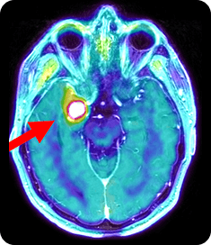
18F-FET/MRI Fusion
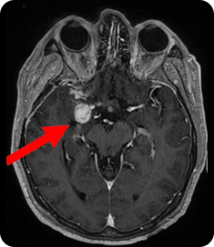
- MRI showed an enlarging enhancing lesion with edema, but whether it was recurrence or radiation necrosis remained inconclusive
- The clarity of 18F-FET revealed a lesion TBRmean of 2.14 and TBRmax of 4.66, further supporting the presence of metabolically active tumor tissue
T1 Post Contrast
18F-FET/MRI Fusion
- MRI showed an enlarging enhancing lesion with edema, but whether it was recurrence or radiation necrosis remained inconclusive
- The clarity of 18F-FET revealed a lesion TBRmean of 2.14 and TBRmax of 4.66, further supporting the presence of metabolically active tumor tissue
These agents are not made commercially available or approved for gliomas. They can be made and used investigationally.
11C-MET, 11C-methyl-L-methionine; CT, computed tomography; EANM, European Association of Nuclear Medicine; EANO, European Association of Neuro-Oncology; 18F-FDOPA, 18F-fluorodopa; 18F-FET, 18F-fluoro-ethyl-tyrosine; MRI, magnetic resonance imaging; PET, positron emission tomography; SNMMI, Society of Nuclear Medicine and Molecular Imaging; TBRmean, mean tumor-to-background ratio.
References: 1. Referenced with permission from the NCCN Clinical Practice Guidelines in Oncology (NCCN Guidelines®) for Central Nervous Center Cancers V3.2025. © National Comprehensive Cancer Network, Inc. 2025. All rights reserved. Accessed March 4, 2026. To view the most recent and complete version of the guideline, go online to NCCN.org. NCCN makes no warranties of any kind whatsoever regarding their content, use or application and disclaims any responsibility for their application or use in any way. 2. Law I, Albert NL, Arbizu J, et al. Joint EANM/EANO/RANO practice guidelines/SNMMI procedure standards for imaging of gliomas using PET with radiolabelled amino acids and [18F]FDG: version 1.0. Eur J Nucl Med Mol Imaging. 2019;46(3):540-557. 3. Galldiks N, Lohmann P, Aboian M, et al. Update to the RANO working group and EANO recommendations for the clinical use of PET imaging in gliomas. Lancet Oncol. 2025;26(8):e436-e447. 4. Data on File. Patient Cases. Telix Pharmaceuticals. 2025.